

Dental Technology
Advanced Dental Technology at Reilly Dental
At Reilly Dental in Marietta, GA, we’re committed to providing exceptional care using the latest advancements in dental technology. Our state-of-the-art tools enhance precision, efficiency, and comfort, ensuring the best possible outcomes for your oral health. From diagnostics to treatment, we combine innovation with expertise to give you a superior dental experience.
Comprehensive Digital Imaging
Digital X-Rays
Digital X-rays allow us to capture detailed images of your teeth and jaw with minimal radiation exposure. These high-quality images provide crucial insights for accurate diagnoses and treatment planning
Intraoral Camera & Chairside Monitors
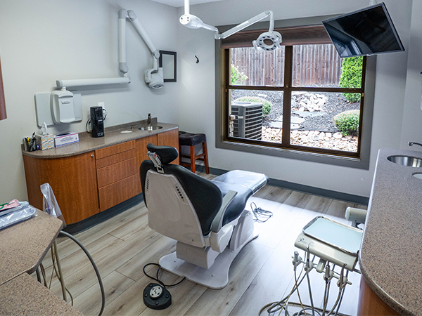
Our intraoral camera provides a close-up view of your teeth and gums, allowing us to identify issues that might not be visible during a routine exam. With chairside monitors, you can see exactly what we see, helping you make informed decisions about your care.
Panorex
The Panorex system creates a comprehensive, 360-degree view of your entire mouth, including teeth, jawbones, and sinuses. This advanced imaging tool is essential for evaluating complex dental concerns, such as impacted teeth or jaw misalignments.
Precision Tools for Better Treatment
Soft Tissue Laser
Our soft tissue laser provides a minimally invasive option for a variety of procedures, including gum contouring and treating periodontal disease. With greater precision, faster healing times, and reduced discomfort, laser technology improves both your experience and results.
Wax-Up Models
Wax-up models are used to plan restorative and cosmetic treatments, such as crowns, veneers, or smile makeovers. These custom models help us visualize and perfect your treatment plan, giving you a preview of your future smile.
ICAM 3D Photogrammetry System
3D dental photogrammetry is a high-precision digital imaging technique used to capture the exact 3D spatial coordinates, positions, and angulations of dental implants , primarily for complex full-arch (All-on-4 / All-on-X) restorations
Four-Camera Architecture
Uses four synchronized moving cameras and a projector instead of a standard two-camera or handheld intraoral scanner setup.
Sub-5-Micron Precision
Triangulates implant positions with aerospace-grade accuracy to guarantee a passive fit for All-on-4 and multi-unit implant cases.
Extraoral Scanning
Captures measurements outside the mouth using specialized titanium scanbodies (ICamBodies), avoiding distortions caused by saliva, soft tissue movement, or stitching errors common in large-span intraoral scans.
Workflow Integration
Combines the photogrammetry implant data with standard soft-tissue intraoral scans, exporting files directly into major CAD software like Exocad or 3Shape.
ICAM 3D Photogrammetry System
3D dental photogrammetry is a high-precision digital imaging technique used to capture the exact 3D spatial coordinates, positions, and angulations of dental implants , primarily for complex full-arch (All-on-4 / All-on-X) restorations
Four-Camera Architecture
Uses four synchronized moving cameras and a projector instead of a standard two-camera or handheld intraoral scanner setup.
Sub-5-Micron Precision
Triangulates implant positions with aerospace-grade accuracy to guarantee a passive fit for All-on-4 and multi-unit implant cases.
Extraoral Scanning
Captures measurements outside the mouth using specialized titanium scanbodies (ICamBodies), avoiding distortions caused by saliva, soft tissue movement, or stitching errors common in large-span intraoral scans.
Workflow Integration
Combines the photogrammetry implant data with standard soft-tissue intraoral scans, exporting files directly into major CAD software like Exocad or 3Shape.
Benefits in Dental Practice Eliminates Remakes
Ensures precise passive seating, which reduces chair time, adjustments, and costly lab remakes
Instead of Panorex, A Planmeca 3D CT
Instead of Panorex, A Planmeca 3D CT is an advanced cone-beam computed tomography (CBCT) dental imaging system used to capture detailed, three-dimensional digital images of the teeth, jawbones, and maxillofacial structures
3D & 2D Imaging
Combines 3D CBCT scans with traditional 2D panoramic, cephalometric, or extraoral bitewing options depending on the model.
Ultra Low Dose™
A proprietary protocol allowing clinicians to capture 3D scans at a radiation dose lower than standard panoramic X-rays.
CALM® Algorithm
An intelligent software feature that automatically corrects and removes artifacts caused by patient movement during the scan.
Versatile Field of View (FOV)
Offers adjustable scan volumes—ranging from small areas focused on a single tooth to large volumes covering the entire craniofacial and ENT (ear, nose, and throat) regions
All Services
Learn More
Keep
In Touch


Managed and Designed by:
